Your cart is currently empty!
Blog
Blueberry (Vaccinium Corymbosum) Blue Crop and Chandler
11.08.2022
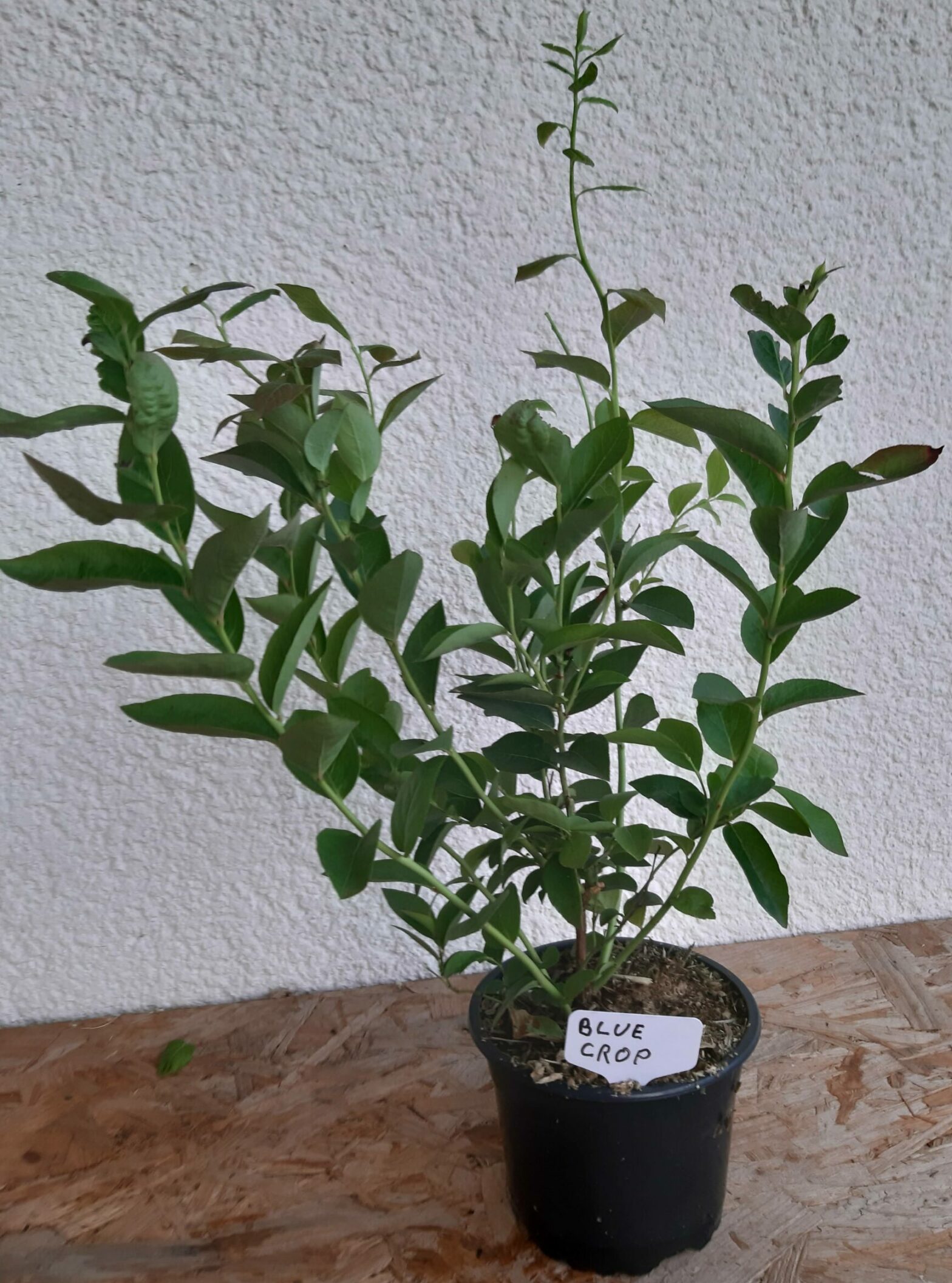
Blueberries are well know to be a highly nutritious berry and an opportunity presented itself to acquire two more varieties (Blue Crop and Chandler) for a very good price…Lidl had them on sale for 3,99€ per plant. Not only were the plants relatively inexpensive but upon examination it was seen that there was several branches that had new growth on them. Thus soft wood was available for cuttings.
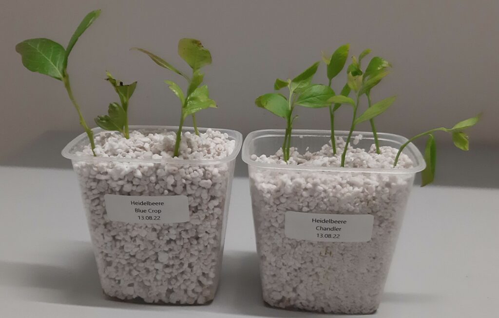
Fig. 1 Blueberry varieties “Blue Crop” (Left) and “Chandler” 13.08.2022
Fig. 2 Cuttings taken from “Blue Crop” (Left) and “Chandler” (Right) Fig. 3 Four cuttings from “Blue Crop” and six cuttings from “Chandler” were taken and set in moistened Pearlite. Sweet Potatoes (Ipomoea batatas)
The motivation for planting sweet potatoes is to avoid the destructive behavior of the Colorado potato beetle (Leptinotarsa decemlineata). In the winter and early spring attempts were made to start sweet potato plants from sweet potatos that were bought in the supermarket. The attempt was of limited success and only a few slips made it to the field. Those that were not attacked by inhabitants of the field did not root and perished.
After being severed from its roots one sweet potato plant was able to be saved and cuttings were made out of the vine. It is hoped that the cutting can be rooted and used as a source for new plants in the 2023 season. Up until the point of it being damaged the sweet potato plant was doing well in the field.
Fig. 1 23.07.2022 Cuttings of sweet potato vines that were recovered from the last surviving damaged plant in the growing bed. Fig. 2 Cutting of Sweet Potato vine in water.
During the period of initial root formation the cuttings of the sweet potato vine will be kept on a window sill that will not receive direct sunlight.
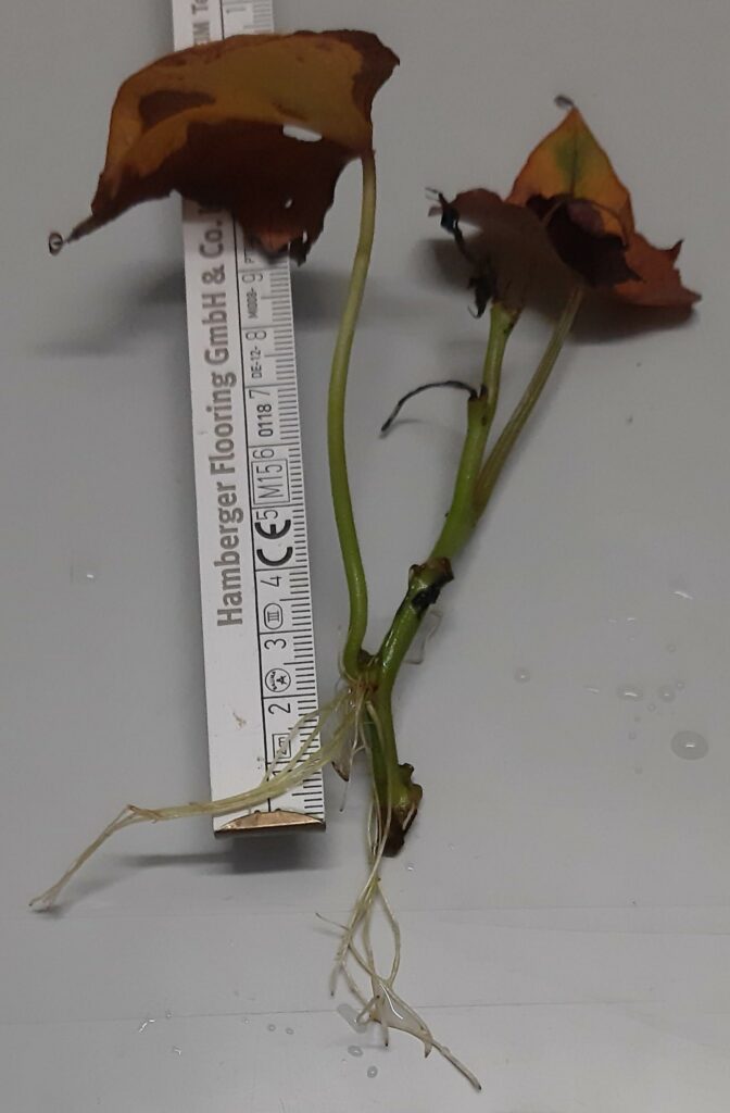
27.07.2022 After three day in water on the window sill the beginnings of root formation was noticed. Three examples of the 7 cuttings are shown below in Fig. 3.
Fig. 3 Three examples of the sweet potato cuttings showing root developement after four day in the water. The appearance of roots so quickly is an unexpected surprise. This result also suggests that it would be much easier to propogate cuttings from a mother plant that has been grown over the winter months.
12.08.22
Six of the seven cuttings have survived. The two smaller cuttings in the glass on the right in Fig. 2 have shown more robust root development than those of the glass on the left.
Fig 4. Sweet potato cuttings from the glass on the right in Fig. 2 after seventeen days in water. Fig. 5 Sweet potato cuttings from glass on the left in Fig 2. The rood developement is not as advanced as for the cuttings in Fig. 4 from the glass on the left. A common element that the four cuttings in Fig. 5 have is that there is more older growth (longer stems) than the two cutting in Fig.4. Also, there was an odor noticed a few days after the cuttings were placed in the water and the water became cloudy much faster than the smaller glass.
When the cuttings were examined more closely after seventeen days. It was seen that the bark had softened and was falling away from the stem when lightly disturbed. The decomposition of the bark could have been the source for the odor that was observed. The segments of the stems that were showing decomposition of the bark were removed from the cuttings.
13.08.2022
Fig. 6 Cuttings A1 and A2 were placed in a mixture of garden soil and potting soil
The two cuttings with advanced root development (call them A1 and A2) were placed in a mixture of garden soil and potting soil. The garden soil carries indigenous microbes and fungi are from environment they will be placed in.
19.08.2022
After seven days it can be seen that the 3 cuttings whose stems were shortened due to partial decomposition began to show robust root growth. Two of these cuttings are ready for planting in soil, name them B1 and B2. The third cutting will be further divided into smaller cuttings. It can be designated C and the cuttings from it C1-4.
12.08.2022 19.08.2022 Fig. 7 comparison of B1 seven days after removal of decomposing stem section. 12.08.2022 19.08.2022 Fig. 8 comparison of B2 seven days after removal of decomposing stem section. 12.08.2022 19.08.2022 Fig. 9 Comparison of cutting C seven days after removal of decomposing stem section. Fig. 10 Cutting C divided into 4 smaller cuttings
01.09.2022
All but the middle segment in the C-group of the cuttings have put forth roots. The cutting that did not continue to grow in the C group was the cuttings taken second from the bottom from the main stem shown in Fig. 10
B1 01.09.2022 B2 01.09.2022 Fig. 11 Cuttings from the B group 01.09.2022. The roots have continued to develope. 01.09.2022 C1 before leaf removal 01.09.2022 C1 after leaf removal FIg. 12 Cutting C1. This was the upper most segment taken from the C-series. The leaves on cutting C1 have turned color and were removed. C2 01.09.2022 C3 01.09.2022 Fig. 13 Cutting C2 was taken from the lowest part of the vine for the C-series and C3 is new growth that sprouted from this lower part. It can be seen that the bottom segment in the C-Series had the least root development. However the new growth that did develop from this section became a cutting on its own, C3.
03.09.2022
A1 03.09.2022 A2 03.09.2022 Fig.14 Plants A1 and A2 on 03.09.2022. The cuttings A1 and A2 that were placed in soil on 13.08.2022 have developed nicely. During their approximately 3 weeks time in the soil they were on a balcony that recieved the late afternoon sun.
Fig. 15 Plants B1 and B2 after transfering to growing containers. Fig. 16 Cuttings in the C-Series after being transfered to the growing containers. Johanniskraut Oil Cream For Irritated And Dry Skin
Manufaktur PUR Johanniskraut and Colloidal Oatmeal Cream is formulated to relieve the symptoms of dry and irritated skin. The Johanniskraut is grown and harvested at the permaculture project being conducted by Manufaktur PUR in Sachsen.
Two components of the cream that provide it with its soothing properties are Johanniskraut oil and colloidal oatmeal. Both of these ingredients have been known to have a positive effect on relieving skin irritation since antiquity. However, it has not been until the development of modern analytical methods that the mechanism responsible for the effects that the ancients have observed have been able to be conclusively explained.
Two bioactive components that are found in Johanniskraut that have been shown to relieve irritation in several studies are Hyperforin and Hypericin [1-5]. These two components along with many other additional secondary metabolites are extracted from the Johanniskraut flowers through a maceration process and the oil is used to make the cream. The traditional method for producing Johanniskraut oil is to place the Johanniskraut in a glass jar filled with olive oil and place the jar in a sunny location, such as a window sill. However, through the research conducted by Heinrich et al., it has been determined that Almond oil is more effective at extracting the secondary metabolites from the plant material than olive oil [6]. In addition, lighting conditions and temperature play a role in the concentration of secondary metabolites that are extracted [7]. The maceration conditions that have been chosen for the production of the johanniskraüt oil are 40°C in a darkened container. According to the data of Heinrich et al., these conditions should have the most favorable effect on the concentration of secondary metabolites in the johanniskrautöl. The increase in temperature to 40°C over that of room temperature has the most significant effect on the hypericin concentration (and increase of approximately 86% ) in the johanniskraütöl compared to the other secondary metabolites that were investigated.[7]
Would healing effects have also been observed when Johanniskraut oil has been applied to areas of the epidermis which have become damaged through various processes [5,8]. Investigation into the mechanism of action has revealed that the wound-healing activity of Hypericum perforatum extract seems to be mainly due to the increase in the stimulation of fibroblast collagen production and the activation of fibroblast cells in polygonal shape which play a role in wound repair by closing damaged area [9,10].
Colloidal Oatmeal has been shown to be effective in relieving the symptoms of epidermal aliments such as xerosis [11], atopical dermatitis [12] and psoriasis [13]. It has been hypothesized that the demonstrated ability of collodial oatmeal to relieve itch and alliviate moderate to severe dry skin while improving the stratum corneum function is attributed to its anti-inflammatory and antioxidant activity [14]. The ability of colloidal oatmeal to be effective against epidermal aliments that are accompanied by irritation appears to be due to its ability to disrupt several processes that lead to inflammation. An investigation of these mechanisms has been conducted by Reynertson et al., and the details are discussed in their publication [14].
In addition to its anti-inflammatory and antioxidant properties, colloidal oatmeal has demonstrated an ability to reduce transepidermal water loss (TEWL). This is due to the presence in oats of the polysaccharides starch and beta-glucan [15,16] which have the ability to retain moisture. When colloidal oatmeal is dispersed in water it deposits fine particles on the skin and forms a viscous occlusive barrier. The polysaccharide beta-glucan exhibits viscosities in solutions that can exceed those of other biological hydrocolloids and significantly contribute to the water-binding properties of oats. The occlusive and water-binding colloidal film holds moisture in the stratum corneum thus improving dry skin conditions [14].
References
1) L. Dellafiora, G. Galaverna, G. Cruciani, C. Dall’Asta, R. Bruni, „On the Mechanism of Action of Anti-Inflammatory Activity of Hypericin: An In Silico Study Pointing to the Relevance of Janus Kinases Inhibition“, Molecules, 23(12), 2018, 3058
2) E. Tedeschi, M. Menegazzi, D. Margotto, H. Suzuki, U. Förstermann, H. Kleinert „Anti-Inflammatory Actions of St. John’s Wort: Inhibition of Human Inducible Nitric-Oxide Synthase Expression by Down-Regulating Signal Transducer and Activator of Transcription-1 (STAT-1 ) Activation“ J Pharmacol Exp Ther., 307(1), 2003, 254-61
3) A. Koeberle, A. Rossi, J. Bauer, F. Dehm, L. Verotta, H. Northoff, L. Sautebin, O. Werz „Hyperforin, an anti-inflammatory constituent from St. John’s wort, inhibits microsomal prostaglandin E2 synthase-1 and suppresses prostaglandin E2 formation in vivo“, Front Pharmacol. 2011 Feb 18;2:7
4) M. Berköz, O. Allahverdiyev, M. Yildirim „Investigation of the effect of Hyperforin and hypericin on inflammatory response in RAW 264-7 Micropflanges“, Van Tip Dre, 25(2), 2018, 124-131
5) U. Wölfle, G. Seelinger, C. M. Schempp „Topical application of St. Johns Wort (Hypericum preforatum)“, Planta Med., 80(2-3), 2014, 109-20
6) M. Heinrich, V. Vikuk, R. Daniels, F. C. Stintzing, D. R. Kammerer, „Characterization of Hypericum perforatum L. (St. John’s wort) macerates prepared with different fatty oils upon processing and storage“ Phytochemistry Letters, 20, 2017, 470-480
7) M. Heinrich, R. Daniels, F. C. Stintzing, D. R. Kammerer, „Comprehensive phytochemical characterization of St. John’s wort (Hypericum perforatum L.) oil macerates obtained by different extraction protocols via analytical tools applicable in routine control“ Pharmazie., 72(3), 2017, 131-138.
8) S. Samadi, T. Khadivzadeh, A. Emami, N. S. Moosavi, M. Tafaghodi, H. R. Behnam, „The Effect of Hypericum perforatum on the Wound Healing and Scar of Cesarean“, J Altern Complement Med., 16(1), 2010, 113-7.
9) N. Oztürk, S. Korkmaz, Yusuf Oztürk, „Wound healing activity of St. Johns Wort (Hypericum perforatum L.) on chicken embryonic fribroblasts“, J Ethnopharmacol., 111(1), 2007, 33-9
10) M. Dikmen, Y. Oztürk, G. Sagratini, M. Ricciutelli, S. Vittori, F. Maggi, „Evaluation of the Wound Healing Potentials of Two Subspecies of Hypericum perforatum on Cultured NIH3T3 Fibroblasts“, Phytother Res., 25(2), 2011, 208-14
11) A. N. Kalaaji, W. Wallo „A randomized controlled clinical study to evaluate the effectiveness of an active moisturizing lotion with colloidal oatmeal skin protectant versus its vehicle for the relief of xerosis“, J Drugs Dermatol. 13(10), 2014, 1265-8. PMID: 25607563.
12) Allais B, Friedman A. „ARTICLE: Colloidal Oatmeal Part II: Atopic Dermatitis in Special Populations and Clinical Efficacy and Tolerance Beyond Eczema“, J Drugs Dermatol.19(10), 2020, s8-s11
13) J.F. Fowler Jr, H. Woolery-Lloyd, H. Waldorf, R. Saini „Innovations in natural ingredients and their use in skin care“, J Drugs Dermatol. (6 Suppl), 2010, S72-81; quiz s82-3
14) K. A. Reynertson, M. Garay, J. Nebus, S. Chon, S. Kaur, K. Mahmood, M. Kizoulis, M. D. Southall, „Anti-Inflammatory Activities of Colloidal Oatmeal (Avena sativa) Contribute to the Effectiveness of Oats in Treatment of Itch Associated With Dry, Irritated Skin“, J Drugs Dermatol. 2015;14(1):43-48.
15) L. Z. Wang and P. J. White. „Structure and physicochemical properties of starches from oats with different Lipid contents“, Cereal Chem. 71, 1994, 443-450
16) A. Ahmad, F. M. Anjum, T. Zahoor, H. Nawaz, Z. Ahmed, „Extraction and characterization of beta-D-glucan from oat for industrial utilization“, Int J Biol Macromol. 46(3), 2010, 304-9.